Analysis of the Abdominal Radiograph
The analysis of the abdominal radiograph taken on the seventh day involves the visual inspection of the X-ray to count the remaining Transit-Pellets radiopaque markers in the colon, as well as the identification of their distribution across the different segments of the colon. By using multiple markers over a period of six days, it eliminates the need to track specific markers from day to day and provides a more comprehensive and accurate measurement of colonic transit.1, 2
It is routine to initially evaluate the overall colonic transit, which is deemed the primary indicator. If the overall transit is abnormal, it is then essential to further assess individual segments to identify the location of any delays within the colon.1
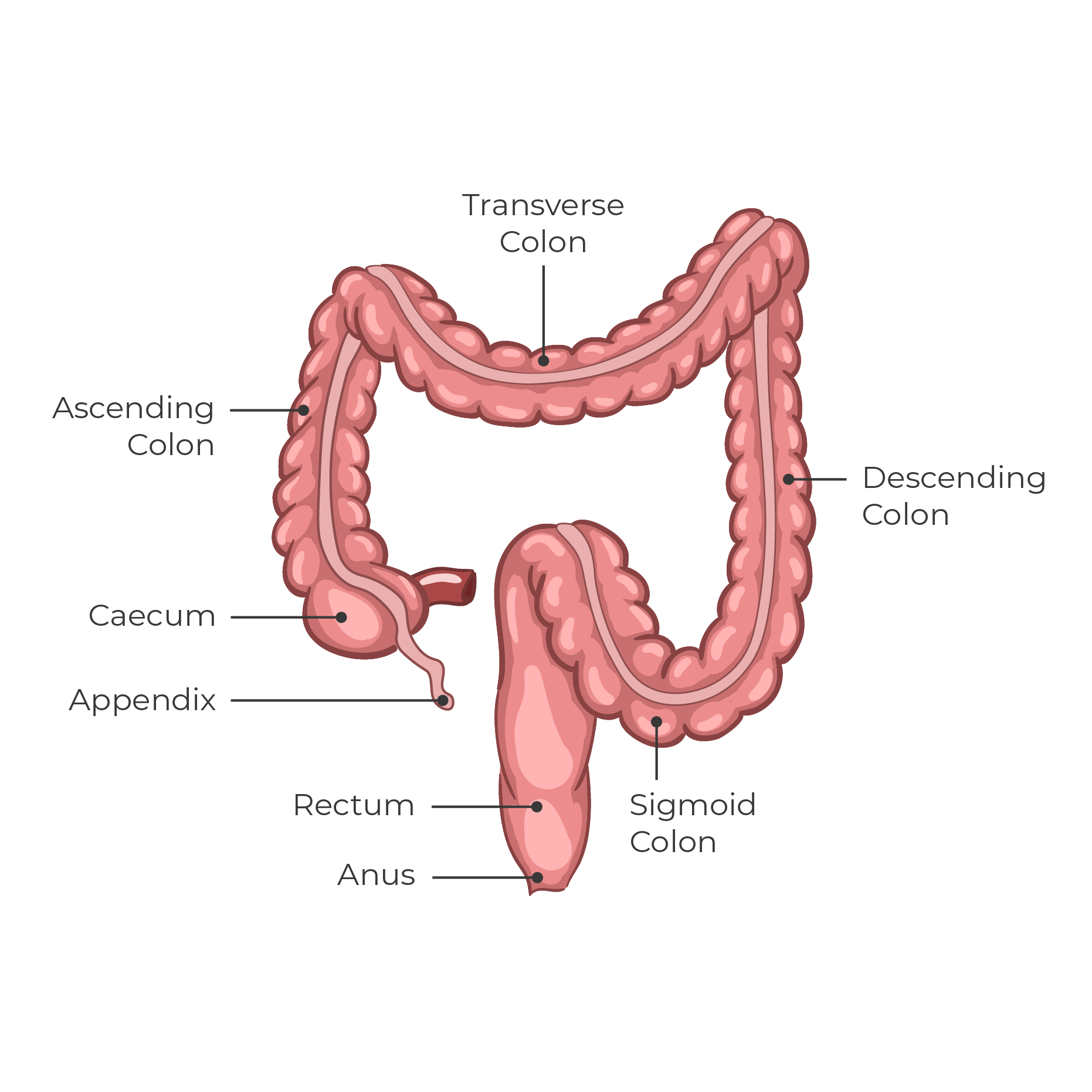
For calculation of segmental transit, four segments are used: caecum-ascending colon, transverse colon, descending colon and sigmoid colon-rectum.1, 2
Some clinicians divide the colon into three regions based on bony landmarks3: the right colon, left colon, and sigmoid colon-rectum. Both approaches are medically equivalent and it´s up to the individual clinic to choose the principle they are most comfortable with.
Calculation of Total and Segmental Transit Time1
Colonic transit is calculated as the mean oro-anal transit time (OATT, mouth-to-anus) for the daily marker doses swallowed. Because the colonic transit constitutes the main part of the mouth-to-anus transit time, OATT is used as a measure of colonic transit. The transit time is equivalent to the number of daily marker doses retained.
When conducting a Transit-Pellets test, it is typically not necessary to differentiate between ring-formed and tube-formed markers. Each marker should be considered as one unit, regardless of its shape. With ten markers per day, each marker is equivalent to 0.1 days (or 2.4 hours).
The formula is straightforward, it involves dividing the number of remaining markers by the daily dose, which in this case is 10 markers. Clinics can use the formula M/10 to determine both total and segmental transit time in days. Some clinics may prefer to present the results in hours, in which case the formula Mx2.4 can be used.
If for example a total of 35 markers are retained, the OATT is determined as 3.5 days (or 84 hours).
Measurement of colonic transit time with radiopaque markers. A Transit-Pellet test in a female patient who ingested 10 markers daily for 6 days. The plain abdominal radiograph taken on day 7 demonstrates that 55 markers are retained. Thus, colonic transit time is 5.5 days. The markers are evenly distributed throughout the colon; the results indicate slow-transit constipation. Upper reference value is 4.0 days for women and 2.2 days for men. (Source: Medifactia).
The Importance of Segmental Transit Time Measurement1
In a clinical practice, it is standard practice to initially assess the overall value, which is considered the most important parameter. Should the overall value be elevated, it is imperative to evaluate individual segments to ascertain the location of any delays within the colon. For example, it can indicate if the dysfunction is localized in the recto-sigmoid area, or if it is more widespread and affects the left colon or even the entire colon, as seen in cases of colonic inertia.
It is also recommended that the segmental transit time be calculated in patients with both rapid transit and diarrhoea. For example, if a patient with diarrhoea has a recto-sigmoid segmental transit time that falls within the upper range of normal transit, it can effectively rule out the possibility of bile acid diarrhoea.4
Why Tube-Formed Markers on Day Six?
The use of tube-form markers on the sixth day serves a specific purpose in certain cases. In patients with slow transit, it may be difficult to accurately locate the caecum. As the tube-formed markers are the last markers to be ingested, their presence in the colon can indicate the location of the caecum. Additionally, a redundant sigmoid colon may overlap with the right colon, while a large caecum can crossover to the rectosigmoid area, the use of tube-formed markers can assist radiologist in identifying the localization of the markers in the correct colonic segment.
The tube-formed markers ingested on day six, approximately 12 hours prior to the X-ray on day seven, can also provide valuable information in cases of rapid transit which is of significance in certain populations experiencing chronic diarrhoea.5
It is also worth noting that if at least six tube-formed markers from the sixth day have not reached the colon by the seventh day, it may be an indication of slow transit in the small intestine, which is a common occurrence in patients with constipation.
Interpretation1
The total number of remaining markers in the colon determines if the colonic transit is delayed or not. A value above the upper reference value (95th percentile) indicates a delay in transit, whereas a value below the lower reference value (5th percentile) indicates an abnormally rapid transit.
For healthy women, the upper reference value for colonic transit time is 4.0 days (40 markers), and a value between 4.1 and 5.0 days is considered moderately delayed transit, while a value greater than 5.0 days is considered clearly delayed transit. On the other hand, healthy men have a more rapid mean transit time than healthy women. The upper reference value for men is 2.2 days (22 markers). Please refer to the tables below for additional guidance.
If a patient has a value below the lower reference value (0.6 days or 6 markers for women and 0.5 days or 5 markers for men), this is considered to be an abnormally rapid colonic transit. Additionally, the distribution of markers in the various colonic segments can provide information about the type of delay. It is, however, important to note that healthy individuals may have a transit value in a few segments that approaches the upper reference value, but not in all segments at the same time, as indicated by the reference value for the total transit time.
A numerical transit time value can be given if the number of retained markers is in the range 3–55 markers. Thus, at least half a daily dose should be excreted and at least half of the evening dose on day six must be retained. If the number of retained markers is only 0–2, the transit time is less than 0.3 days. If 56–60 markers are retained, the transit time is more than 5.5 days. This is an indication that equilibrium has not been reached yet.
The tables below provide a reference for the evaluation of total and segmental colonic transit. These values may be used as a guide for interpretation of the results.
Result in Days
Colonic Transit Time (OATT); Reference Values 2, 5, 6
Normal transit time corresponds to the range from percentile 5 to percentile 95 in the control material. Reference values based on 199 subjects.
Segmental Transit Time; Upper Reference Values 2
Percentile 95 calculated per segment in healthy subjects.
For clinics that prefer to obtain results in hours, please click HERE.
Guidance on reference values for colonic transit time in children and adolescents, please click HERE.
- 0020 Clinical Evaluation Report – Transit-Pellets ver08 (Internal Document). November 2023.
- Abrahamsson, H., Antov, S. & Bosaeus, I. (1988). Gastrointestinal and colonic segmental transit time evaluated by a single abdominal X-ray in healthy subjects and constipated patients. Scand J Gastroenterol. Vol. 23 (suppl 152), 72-80.
- Arhan P., Devrode G., Jehannin B., et al (1981). Segmental colonic transit time. Dis Colon Rectum, Vol. 24(8), 625-629.
- Sadik, R., Abrahamsson, H., Ung, K.A., et al. (2004). Accelerating regional bowel transit and overweight shown in idiopathic bile acid malabsorption. Am. J. Gastroenterol., Vol. 99, 711-718.
- Sadik, R., Abrahamsson, H., Stotzer, P.O. (2003). Gender differences in gut transit shown with a newly developed radiological procedure. Scand. J. Gastroenterol., Vol. 38, 36-42.
- Törnblom, H., et al. Data on File, Gastrointest Lab, Sahlgrenska University Hospital.


